Redness in skin folds
Distinguishing Psoriasis, Tinea, Candida, and Contact Dermatitis for Redness in Armpits and Groin
For sensitive areas, to maintain anonymity Lesion take photos of only the boundaries, and record sweat, friction, hair removal, deodorant,ointment Record before and after use.
Click on unfamiliar terms to see their meanings immediately.
Psoriasis in skin folds may appear smooth and red with less scaling due to moisture. Look for ring edges or small surrounding pustuleareas; if a boundary where the product was applied is visible, check for tinea, candida, or contact dermatitis.
Points to check in the photos first
Even if they look similar, the dead skin, blisters, and borders may differ
We have provided both typical appearances and examples that look slightly different. Do not decide based on a single similar photo; compare how long symptoms like dead skin, wheals, comedones, or oozing have persisted in the same area.
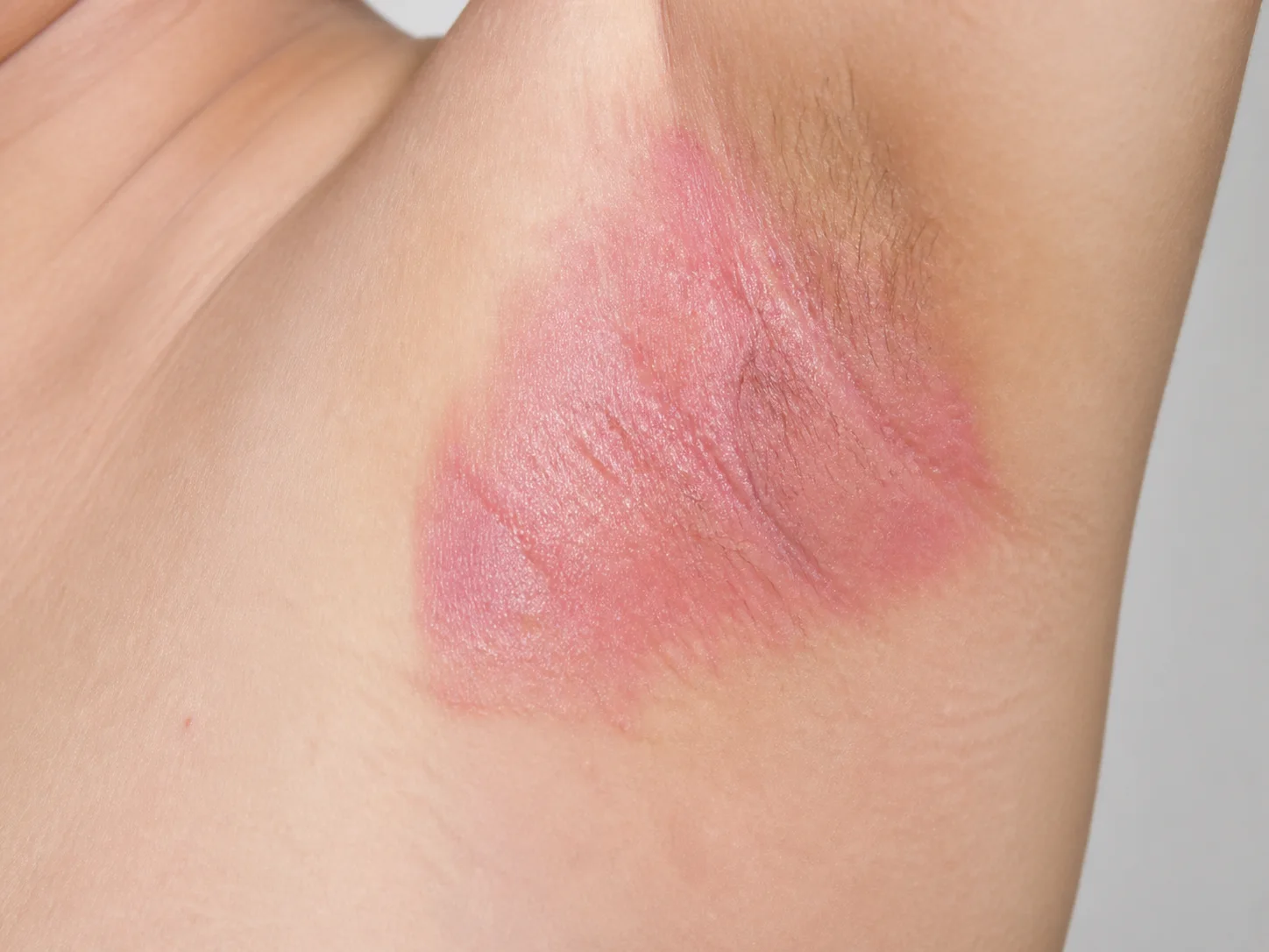
In moist skin folds, psoriasis may have fewer thick scales and can resemble candidiasis, tinea, or friction dermatitis. We examine the borders and look for clues of psoriasis in other areas.
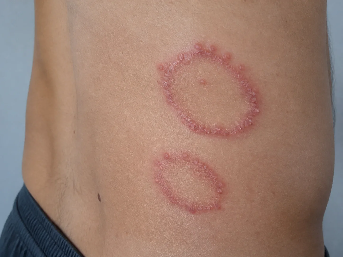
Tinea corporis is characterized by a ring-shaped pattern where redness and scaling are more active on the outer edge than in the center. We confirm the need for infection treatment after differentiating it from nummular eczema or pityriasis rosea.
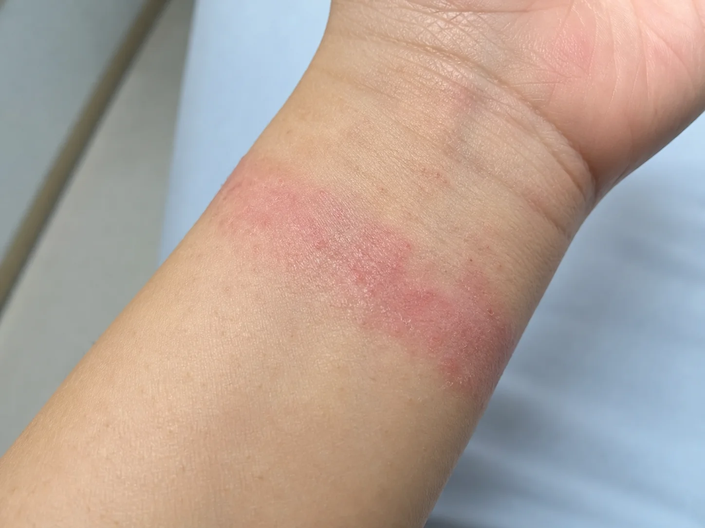
Even if the boundary is not perfectly straight, whether the rash overlaps with the area of contact, such as from a watch, band, or adhesive, serves as a clue. The date the product was changed is also verified.
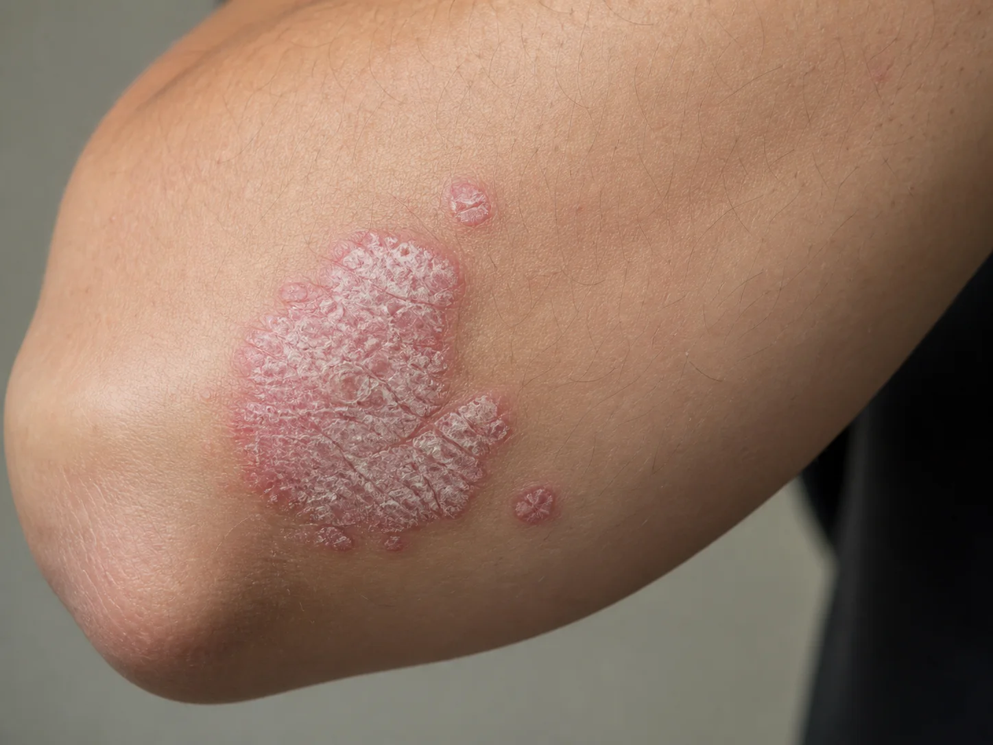
Psoriasis is evaluated by looking at the borders of the plaques, the layered scales, and the distribution, such as on the elbows and knees. Changes in the nails and joint discomfort are also checked separately from the skin photos.
| Points to check | Inverse Psoriasis (Skin Folds) | Tinea | Candidal Intertrigo | Contact Dermatitis |
|---|---|---|---|---|
| Surface | Smooth, shiny red plaques with distinct boundaries; may have little scaling | Rings and scales with a more active outer edge | Moist, bright red color; possible small satellite papules or pustules nearby | Stinging, itching, oozing, or cracking associated with the area of contact |
| Distribution | Relatively symmetrical on both sides of the skin fold; may be accompanied by psoriasis in other areas | Dominant on one side or expanding in a ring shape | Lesions on moist, touching surfaces and surrounding areas | Boundaries where hair removal agents, deodorants, adhesives, or cleansers were applied |
| Things to check together | Navel, behind ears, scalp, nails | Athlete's foot, nails, family contact | Diabetes, antibiotics, immunosuppression, and erosion | Start date of new products and repeated exposure |
| Verification | Examination and clues of psoriasis in other areas | Fungal test if necessary | Examination and fungal confirmation if necessary | Discontinue exposure; patch test if necessary |
Do not rule out psoriasis just because there is little scaling
Skin folds are humid and subject to frequent friction, so typical thick silvery-white scales may be less visible. Check for symmetry on the opposite side and clues on the scalp, navel, and nails.
Observe small dots around the lesion and the outer borders
Satellite lesions of Candida, active edges of tinea, and product exposure boundaries are easily missed when only a wide red plaque is photographed. Capture the center, border, and surrounding normal skin in one shot.
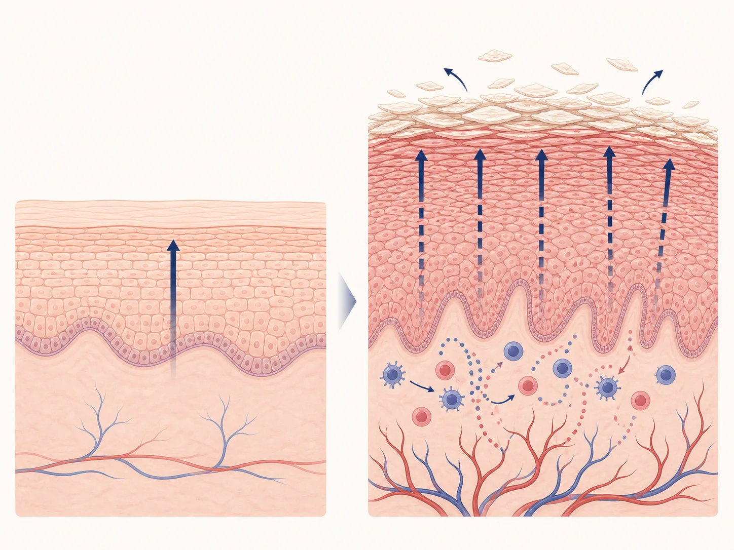
This is a simplified concept showing how overactivation of the immune system contributes to the rapid increase of skin cells, inflammation, and the accumulation of scales.
Check for infection first if there is pain, pus, or rapid spreading
If skin folds are severely painful, swollen, or accompanied by pus, odor, or fever, infection evaluation takes priority over psoriasis self-management.
For sensitive areas, photograph only the necessary range and list the products used
Please take photos showing whether both sides are similar and if there are small pustules around the red area. Also, let us know when you used hair removal products, deodorants, cleansers, or ointments.
Record bilateral symmetry, the outer border of the plaque, surrounding small pustules, and the timing of hair removal, product, or ointment use.
Next reading
Content to follow
If my armpits or groin are smooth and red, is it inverse psoriasis?
How is it distinguished from tinea, candidiasis, or contact dermatitis?
How does herbal medicine treatment categorize psoriasis in humid areas with frequent friction?
Skin terms used in this differential guide
When herbal medicine treatment is the primary focus
We set the goals for herbal medicine treatment based on your previous treatments and when symptoms recurred
Psoriasis can follow a chronic course where skin changes improve and then worsen again, and the burden varies by area, such as the scalp, nails, or skin folds. While continuing existing treatments, herbal medicine treatment can become the primary focus to manage the recurring patterns and overall systemic condition together.
We ask where it appeared and when it worsened again
We examine the redness and thickness of the scales, the distribution of skin changes, sensations of heat or dryness, and symptoms related to sleep, digestion, fatigue, and joints.
We check if not only the itching, but also sleep and daily life are improving
We compare the occurrence of new skin changes, scaling and cracking, nail changes, joint discomfort, and daily functional abilities.
We set the goals for herbal medicine treatment by identifying the problems that need to be resolved first
If there are causes that require immediate priority, such as infection or rapid worsening, those treatments come first. Subsequently, for herbal medicine treatment, we determine the primary goal of recovery by examining current discomfort, recurring patterns, sleep, digestion, and fatigue, and coordinate the sequence with necessary dermatological treatments.
While reducing aggravating factors through lifestyle management, herbal medicine treatment determines the initial target by analyzing the pattern of lesions, sleep, digestion, and fatigue through pattern identification (Byeonjeung). Necessary skin treatments are continued in accordance with the progress.
Consult about treatmentEvidence and Update Information
This content was organized by Korean medicine doctor Choi Yeon-seung and updated on 2026-08-23.
- DermNet, Flexural psoriasis
- DermNet, Psoriasis of the palms and soles
- American Academy of Dermatology, Psoriasis: Signs and symptomsPlaque, guttate, pustular, and erythrodermic psoriasis, and nail/joint symptoms