Differential Diagnosis of Hand Rash
Dyshidrosis, Tinea Manuum, and Contact Dermatitis: How to Distinguish Blisters and Scaling on the Hands
Take close-up photos of the hands only, but record the difference between both hands, foot symptoms, and the date of exposure outside the photo.
Click on unfamiliar terms to see their meanings immediately.
Deep on the sides of the fingers small blisters, a wide area of one palm Keratin, and redness that follows the area of contact are different clues. Please also note the timing of water work, glove use, athlete's foot, or the use of new products or cosmetics.
Points to check in the photos first
Even if they look similar, the dead skin, blisters, and borders may differ
We have provided both typical appearances and examples that look slightly different. Do not decide based on a single similar photo; compare how long symptoms like dead skin, wheals, comedones, or oozing have persisted in the same area.
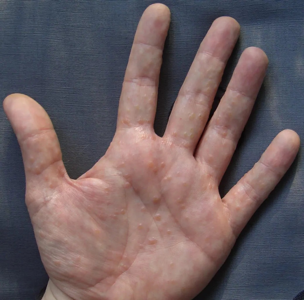
Palms, sides of fingers, small and deep blisters, itching. Differentiate from athlete's foot and contact dermatitis
Photo: Inwe~commonswiki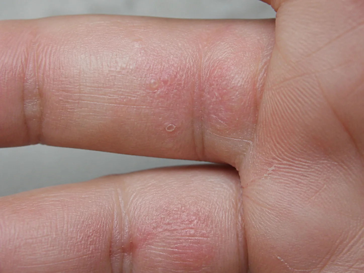
Small, deep blisters on the sides of the fingers or palms are important clues for pompholyx. Since it can overlap with athlete's foot or contact dermatitis, we examine whether it is severe on only one side and consider exposure factors.
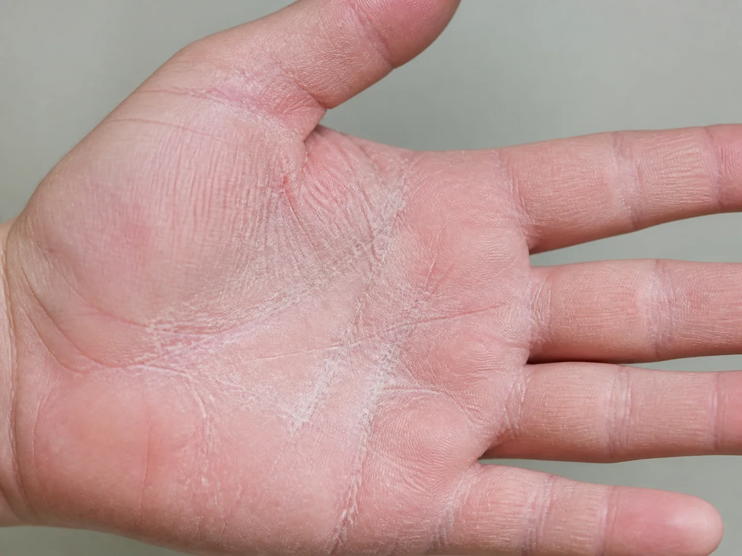
Tinea manuum can appear as fine scaling that is more severe on one hand, which can be confused with hand eczema. We consider accompanying athlete's foot and fungal tests together.
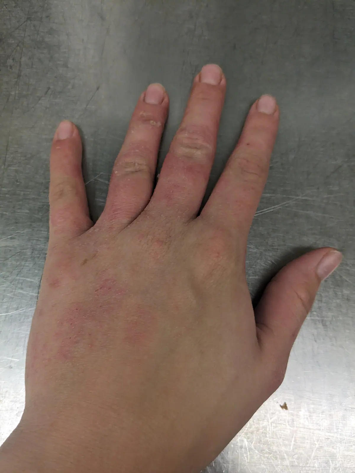
Area of contact · redness and dryness · before and after using a new product. Differential diagnosis from hand eczema and infections.
Photo: Hannah Clover| Points to check | Dyshidrosis | Tinea manuum | Contact Dermatitis |
|---|---|---|---|
| Core Appearance | Deep, small blisters on the sides of fingers and palms | Fine scaling and white powder along skin lines | Redness, stinging, blisters, cracking |
| Distribution | Can occur on both hands; sides of fingers are a clue | One hand may be more severe | May overlap with the area of contact |
| Things to check together | Sweat, stress, season | Athlete's foot / fingernails and toenails | Detergents, gloves, metals, adhesives |
| Verification | Progress and examination | Fungal test if necessary | Removal of exposure / patch test if necessary |
Check if deep, small blisters started on the sides of the fingers
Dyshidrosis is characterized by deep, small blisters on the sides of the fingers and inside the palms, and severe itching, rather than large blisters that burst immediately on the surface. itchingThis is a key clue. Take photos in chronological order, from the appearance of blisters to the stage where the skin peels after the blisters dry.
If keratinization occurs on both one hand and the feet, check for tinea
Tinea manuum can appear only as broad, fine keratinization on one palm, which can be confused with chronic hand eczema. If athlete's foot or changes in fingernails and toenails are present, confirming infection, including a fungal culture test, is the priority.
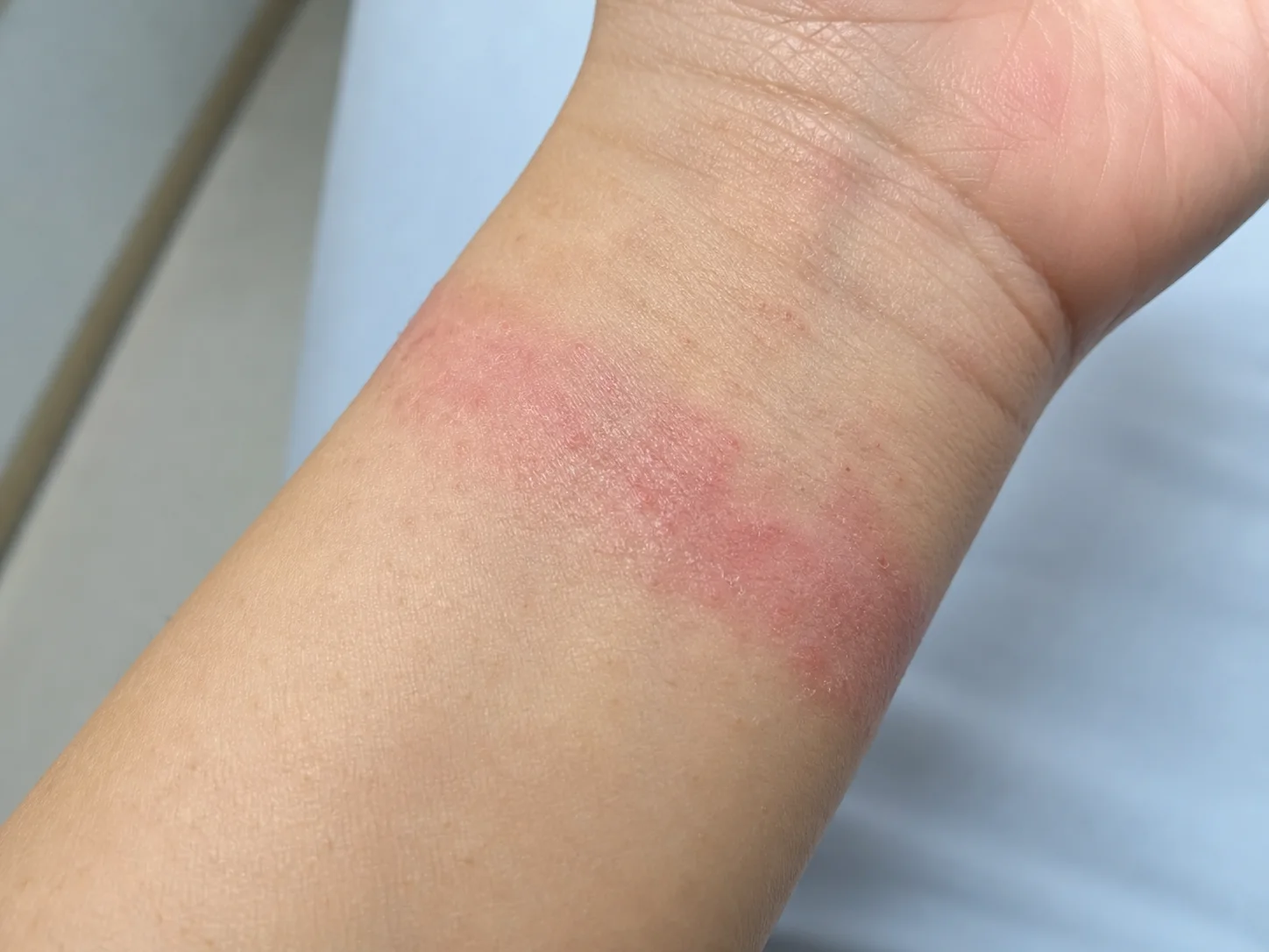
Even if the boundary is not perfectly straight, whether the rash overlaps with the area of contact, such as from a watch, band, or adhesive, serves as a clue. The date the product was changed is also verified.
The area of contact and the date you changed products are clues for contact dermatitis
Check if the rash boundaries overlap with areas of repeated contact, such as the inside of gloves, watches, bands, cleansers, or nail products. As time passes, the boundaries may blur, so photos from the onset and the work environment provide clearer clues.
Please take photos of both hands and feet together
Please take photos of the blisters on the sides of the fingers, keratinization on one hand, the skin between the toes, and changes in the toenails together. Also, please note the dates you started working with water, using gloves, or using new cosmetics.
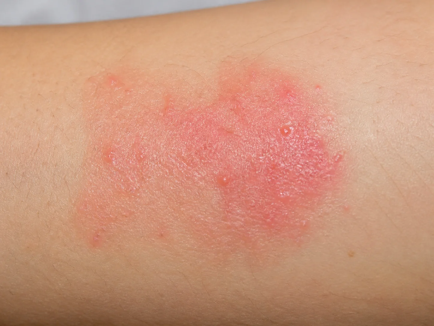
During the acute phase, not only redness but also small blisters, oozing, and peripheral scaling may be visible. If pus, heat, or pain overlap, we first check for infection.
Compare the starting location of small blisters, differences between both hands, athlete's foot, and exposure to water, gloves, or products all at once.
Next reading
Content to follow
If deep, small blisters appear on the hands and feet, is it dyshidrotic eczema?
How can I distinguish dyshidrotic eczema from athlete's foot or contact dermatitis?
Why is herbal medicine treatment important when blisters and cracking recur?
Skin terms used in this differential guide
When herbal medicine treatment is the primary focus
We set the goals for herbal medicine treatment based on your previous treatments and when symptoms recurred
If the cycle of deep blisters on the hands and feet drying, peeling, and cracking repeats, managing only the affected area may leave a burden on your daily life. By dividing the changes into the vesicle stage and the dryness/fissure stage, the recurrence cycle can be managed, centered on Korean herbal medicine treatment.
We ask where it appeared and when it worsened again
We examine the depth of the vesicles, feelings of heat, oozing, subsequent dryness and fissures, as well as sweat, stress, sleep, and the timing of recurrence.
We check if not only the itching, but also sleep and daily life are improving
We compare the interval at which new vesicles appear, the degree of cracking and pain, limitations in the use of hands and feet, and the recovery period.
We set the goals for herbal medicine treatment by identifying the problems that need to be resolved first
If there are causes that require immediate priority, such as infection or rapid worsening, those treatments come first. Subsequently, for herbal medicine treatment, we determine the primary goal of recovery by examining current discomfort, recurring patterns, sleep, digestion, and fatigue, and coordinate the sequence with necessary dermatological treatments.
While reducing aggravating factors through lifestyle management, herbal medicine treatment determines the initial target by analyzing the pattern of lesions, sleep, digestion, and fatigue through pattern identification (Byeonjeung). Necessary skin treatments are continued in accordance with the progress.
Consult about treatmentEvidence and Update Information
This content was organized by Korean medicine doctor Choi Yeon-seung and updated on 2026-08-23.